


- Conventional Radiography is 2-D imaging modality
- It is required to perform minimum 2-views orthogonal to each other:
- 1 AP (Anterior to Posterior) or PA (Posterior to Anterior)
- 2 Lateral
- Supplemental views: Oblique views etc.
- Skeletal radiographs typically use AP & lateral views
- Chest radiographs and Scoliosis imaging in children will usually use the PA technique
- Exceptions for PA chest views: patients unable to cooperate (severely ill or unconscious patients)
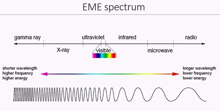
- X-rays are a form of electromagnetic energy (EME) similar to light photons or other sources
- X-rays are a form of man-made radiation
- Ionizing effect of x-rays process of removal of atomic electrons from their orbits
- Two basic types of ionizing radiation:
- Particle (particulate) radiation produced by alpha & beta particles that are the result of radioactive decay of different materials
- Electromagnetic Radiation (EMR) produced by x-rays or gamma rays called photons
- The energy of EMR depends on its wavelength
- Shorter wavelength corresponds to higher energy
- The energy of EME is inversely related to its wavelength

X-ray Properties
- No charge
- Invisibility
- Penetrability of most matters (esp. human tissues) depends on "Z" (atomic number)
- Making compounds fluoresce and emit light
- Travel at the speed of light
- Ionization and biologic effect on living cells
The Imaging System
- X-rays are produced by an imaging system ( x-ray tube, operator's console, and high voltage generator)
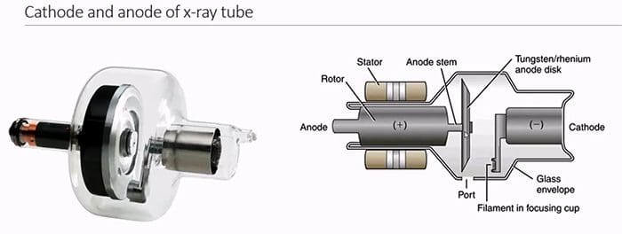
- X-ray tube composed of (-) charged cathode and (+) charged anode enclosed in the evacuated class envelope and housed in the protective coat of metal
- A Cathode made up of filament wire embedded within the focusing cup to give electrostatic focus to electrons' cloud
- Filament wire of heat resistant thorium tungsten metal of high melting point (3400 C) that "boils off" electrons during thermionic emission
- Focusing cup polished nickel (-) charged that accommodated the filament to electrostatically repulse the electrons and confines them to the focal spot of the anode disc where x-rays are produced
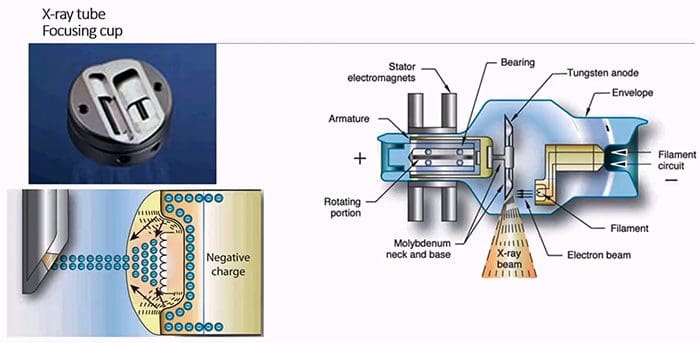
- Anode (+) charged target for electrons to interact at the focal spot
- Conducts electricity
- Rotates to dissipating heat
- Made of tungsten to resist heat
- Anode has a high atomic number to produce x-rays of very high efficiency at the focal spot
- There are 2-focal spots large and small, each corresponding to cathode's filament size (small vs. large) that depends on the magnitude of current in the cathode dictated by a radiographic study of larger or smaller body parts
- It is known as the dual focus principle
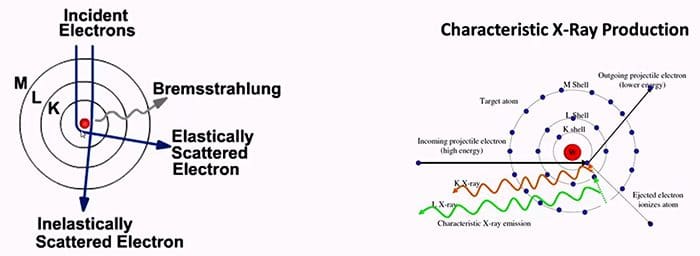
When Electrons are emitted from the cathode as the cloud, they slam into the Anode's focal spot resulting in 3 man events
- Production of heat (99% outcome)
- Production of Bremsstrahlung (i.e., breaking radiation) x-rays that represent the majority of x-rays within the x-ray emission spectrum
- Production of Characteristic x-rays very few in the emission spectrum
- Newly formed x-rays at the anode are of different energies
- Only need high energy or "hard" x-rays to perform the radiographic study
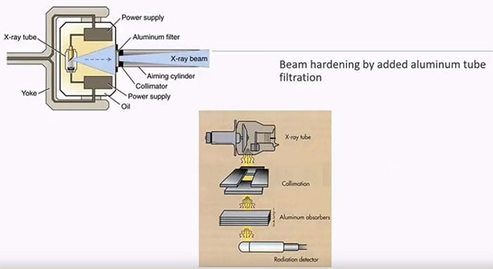
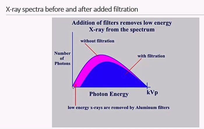
- Before x-rays exiting the tube we need to remove weak or low energy photons, i.e., "harden the beam."
- Added tube filtration in the form of aluminum filters is used that removes at least 50% of the "unfiltered" beam thus minimizing the patient's radiation dose and maximizing image quality
High Voltage Generator
- X-ray production requires an uninterrupted flow of electrons to the anode
- Regular electricity supplies AC power with sinusoidal currents of "peaks and drops."
- In the past, single-phase high voltage generators would convert AC power into a half, or full wave rectified supply with a measure in the thousands of volts delivered with a "voltage ripple" or peaks of high voltage. Therefore, a term kilo voltage peaks (kVp) was used
- Modern generators provide "uninterrupted" flow of electrical potential to the x-ray tube eliminating "voltage ripples" thus referred to as kilovoltage kV without "peaks."
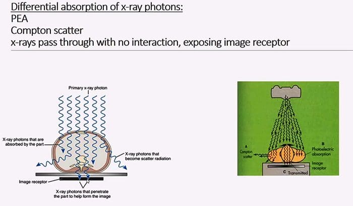
When x-rays interact with the patient's tissued 3 events will occur
- X-rays will pass through without interaction and "expose" the image receptor
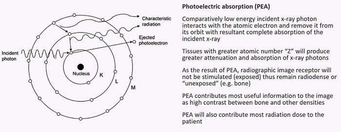
- Photoelectric interaction/effect (PE) comparatively lower energy x-rays will be absorbed/attenuated by the tissues
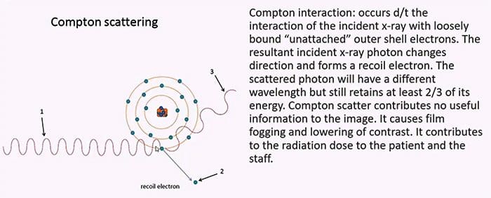
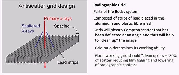
- Compton scatter x-rays are "bounced off" to form scatter, contributing no useful information to the film and lower image contrast while potentially giving unnecessary radiation dose to staff
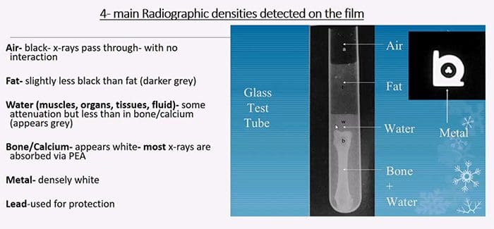
- The final image is the product of all three types of interactions known as
- Differential absorption of x-ray photons - the result of photons' absorption via PE, Compton scatter and x-rays passing through the patient
- Compton scatter probability decreases with an increase in x-ray energy compared to PE effect
- Compton effect probability does not depend on the atomic number (Z)
- An increase of total mass density (thick vs. thin) will increase Compton and PE interaction
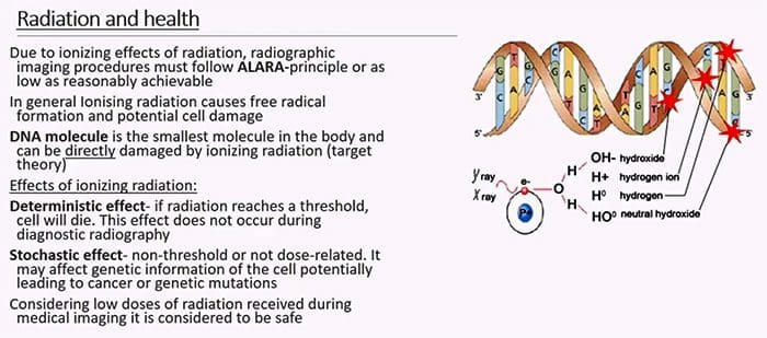
What cells in the body are considered most vulnerable and most resistant to radiation?
- Cells that are rapidly dividing and not terminally differentiated, epithelial cells, etc. are more radiosensitive
- Bone marrow cells (stem cells) & lymphocytes are very radiosensitive
- Muscle & and nerve cells are terminally differentiated and are less sensitive to radiation
- Aged (senescent cells) vs. immature fetal cells are more vulnerable to radiation
- However, following low dose radiation in most healthy individual cells will be able to repair likely without any long-lasting changes

- Pregnancy & radiation initial 6-7 weeks are the most vulnerable
- Do not use routine (non-emergent) radiographic examinations in pregnancy
- Apply 10-day rule establish that radiographs can only be obtained during the initial ten days from the onset of the last menstrual cycle
- Radiographic imaging of children:
- If clinically possible use non-ionizing forms of medical imaging (e.g., ultrasound)

Non-axial imaging studies that use x-ray photons:
- Conventional radiography
- Fluoroscopy
- Mammography
- Radiographic angiography (currently less often used)
- Dental imaging
- Cross-sectional imaging using x-ray photons: Computed Tomography
Indication and Contraindication for conventional radiographic imaging
- Advantages of Radiography: widely available, inexpensive, low radiation burden, the first step in imaging investigation of most MSK complaints
- Disadvantages: 2D imaging, relatively lower diagnostic yield during an examination of soft tissues, numerous artifacts, and dependence on correct radiographic factors selection, etc.
Indications:
- Chest: initial assessment of lung/intrathoracic pathology. Potentially determines or obviates the need for chest CT scanning. Pre-surgical evaluation. Imaging of pediatric patients due to extremely low radiation dose.
- Skeletal: to examine the bone structure and diagnose fractures, dislocation, infection, neoplasms, congenital bone dysplasia, and many forms of arthritis
- Abdomen: can assess acute abdomen, abdominal obstruction, free air or free fluid within the abdominal cavity, nephrolithiasis, evaluate placement of radiopaque tubes/lines, foreign bodies, monitor resolution of postsurgical ileus and others
- Dental: to asses common dental pathologies
