


Serratus anterior is an important muscle for the overhead athlete. Dysfunction in this muscle can lead to shoulder injuries such as impingement, rotator cuff breakdown and performance decrements during overhead tasks. Chiropractor, Dr. Alexander Jimenez looks at its anatomy and biomechanics, and highlights some clinically relevant exercises designed to retrain serratus anterior function.
Shoulder pain is a common complaint in overhead athletes involved in sports such as swimming, tennis and the throwing sports. Overhead upper extremity movements place incredibly high demands on the shoulder complex, requiring high muscular activation around both the scapula-thoracic joint and glenohumeral joint. Researchers have reported that abnormal biomechanics of the shoulder girdle and repeated overhead movements can lead to injuries in overhead throwing athletes(1).
In particular, muscular imbalances around the shoulder complex in the form of altered activation patterns and inherent myofascial restrictions, may lead to diminished scapular control and dyskinesis resulting in glenohumeral joint injuries, such as instability and impingement(2).
The serratus anterior (SA) is one of the scapula muscles that provides a link between the shoulder girdle and the trunk and has often be implicated as a dysfunctional muscle in shoulder pathologies(3,4). The SA is a prime mover of the scapula, contributing to the maintenance of normal scapulohumeral rhythm and motion(4). It has large moment arms to produce upward rotation and posterior tilting due to its insertion on the inferior and medial border of the scapula. Poor activation of the SA muscle may result in reduced scapular rotation and protraction, resulting in relative anterior-superior translation of the humeral head in relation to its glenoid articulation, causing subacromial impingement and rotator cuff tears(5).
Anatomy & Biomechanics
The SA is a flat sheet of muscle originating from the lateral surface of the first nine ribs (see Figure 1). It passes posteriorly around the thoracic wall before inserting into the anterior surface of the medial border of the scapula(6). Overall, the main function of the SA is to protract and rotate the scapula, keeping it closely opposed to the thoracic wall allowing optimal positioning of the glenoid fossa for maximum efficiency for upper extremity motion(7). The SA can be broken down into three functional anatomical components(8,9):
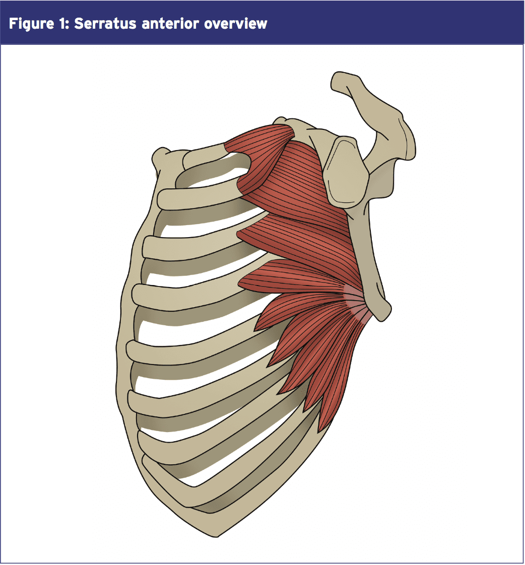
- The superior component originates from the first and second ribs and inserts into the superior medial angle of the scapula. This component serves as the anchor that allows the scapula to rotate when the arm is lifted overhead. These fibres run parallel to the 1st and 2nd rib;
- The middle component of the SA originates from the second, third and fourth ribs and inserts onto the medial border of the scapula anteriorly (sandwiched between the scapula and ribs). This component is the prime protraction muscle of the scapula;
- The inferior component originates from the fifth to ninth ribs and inserts on the inferior angle of the scapula. The fibres form a ‘quarter fan’ arrangement, inserting onto the inferior border of the scapula. This third portion serves to protract the scapula and rotate the inferior angle upward and laterally. Inman (1944) proposed that the lower part of the serratus anterior is the stabiliser of the inferior border of the scapula, and works with the lower trapezius to create a force couple to upwardly rotate the scapula during overhead movement(10).
- Upwardly rotate the scapula during
shoulder abduction, particularly from 30 degrees of shoulder abduction onwards; - Stabilise and protract the scapula during shoulder flexion movements;
- Rotate the inferior angle anteriorly (posterior tilt of the scapula);
- Stabilise the scapula against the thorax during forward pushing movements in order to prevent the scapula ‘winging’ (see below);
- Hold the medial border of the scapula firmly against the thorax so that with the hand fixed, it can displace the thorax posteriorly during a push up.
- Throwing a punch in boxing to achieve maximum reach of the arm. Hence the SA is often referred to as the ‘boxers muscle’.
- In the boxer, the SA is needed to brace the scapula on impact with the punch. This allows maximum transfer of force from the lower limbs into the torso then for it to be imparted into the upper limb and punch. If the scapula was to ‘collapse’ into retraction upon impact of the punch, the boxer would then lose power in the punch.
- In swimming, the swimmer needs full upward rotation to achieve maximum reach in the water upon hand entry in freestyle and butterfly.
- The overhead athlete such as a tennis player needs full upward rotation in the act of serving.
- The sweep style rower needs full protraction on the ‘long’ side to achieve necessary reach during the catch phase of the rowing stroke.
- In baseball, the pitcher needs high levels of protraction during the follow through of the baseball pitch. Similarly in the throwing events in athletics.
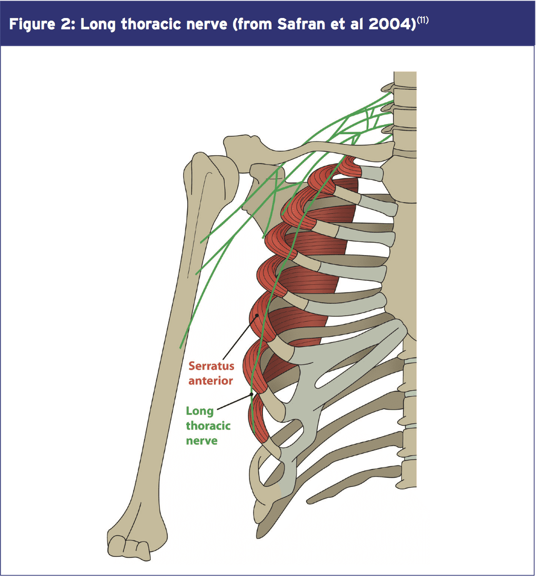
The SA is innervated by the long thoracic nerve, originating from the anterior rami of the fifth, sixth, and seventh cervical nerves (see Figure 2)(7,8). Branches from the fifth and sixth cervical nerves pass anteriorly through the scalenus medius muscle before joining the seventh cervical nerve branch that courses anteriorly to the scalenus medius. The long thoracic nerve then dives deep to the brachial plexus and the clavicle to pass over the first rib. Here, the nerve enters a fascial sheath and continues to descend along the lateral aspect of the thoracic wall to innervate the SA muscle.
SA Dysfunction Associated With Scapula Dyskinesis
Proper positioning of the humerus in the glenoid cavity, known as scapulohumeral rhythm, is critical to the proper function of the glenohumeral joint during overhead motion. A disturbance in normal scapula movement may cause inappropriate positioning of the glenoid relative to the humeral head, resulting in injury such as impingement and instability(2,12,13). Precise timing of muscle activation and adequate levels of muscle recruitment are also needed to position the scapula in the ‘ideal’ position. Small changes of activation in the muscles around the scapula can affect its alignment, as well as the forces involved in upper limb movement(14). One of the primary muscles responsible for maintaining normal rhythm and shoulder motion is the SA(15).Clinically, it has been shown that if a therapist actively repositions a patient’s scapula into an ‘ideal’ posture by reducing the anterior tilt, then it is noticed that impingement pain is reduced, and strength increases in the shoulder during overhead activities(16). The SA is a muscle that will actively work to position the scapula into posterior tilt during overhead activities.
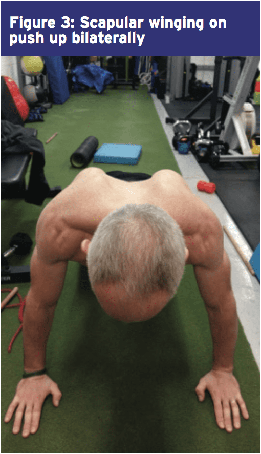
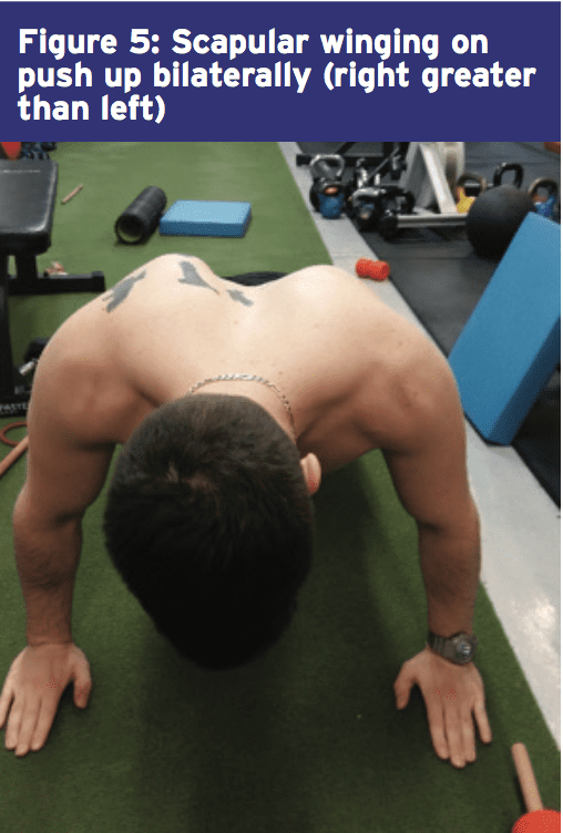
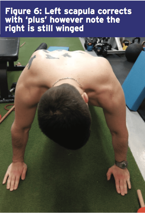
Lack of strength or endurance in the SA allows the scapula to rest in a downwardly rotated and anterior tilted position, causing the inferior border to become more prominent. Furthermore, gross pathological inhibition of the SA or an imbalance between the SA and the other protracting muscle, the pectoralis minor, may result in a ‘winging scapula’. Scapular winging may precipitate or contribute to persistent symptoms in patients with orthopaedic shoulder abnormalities(17,18).
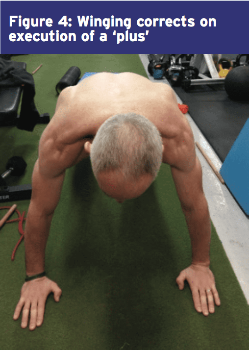
This scapular winging is best appreciated on watching the scapula posture during a push up exercise. Often if the winging is due to a muscle imbalance and the primary scapula stabiliser is the pectoralis minor, this will usually correct if the patient is asked to ‘plus’ and protract the scapula. If the wing disappears then the cause is most likely muscle imbalance, if it remains then it may be a pathological inhibition of the SA. Examples of this are shown below in figures 3-6.
The gross examples of scapular winging can also be due to a pathological lesion to the long thoracic nerve that innervates the SA muscle. For the purposes of this discussion, direct nerve insults to the long thoracic nerve will not be discussed as often these injuries will seriously curtail athletic participation in an athlete. The reader is directed to references 19-23 for a more detailed discussion on these pathological nerve lesions.
SA Importance
The importance of a conditioned serratus anterior muscle has been highlighted in EMG studies of sports such as swimming(24), throwing(25), and tennis(26). A fatigued serratus anterior muscle will reduce scapular rotation and protraction and allow the humeral head to translate anteriorly and superiorly, possibly leading to secondary impingement and rotator cuff tears(27). More direct studies on the role that SA plays in shoulder pathologies has been studied by other researchers. The pertinent points of some of these studies can be summarised below;1. When the trapezius and SA EMG is investigated in people suffering from shoulder pathology is compared with those without pathology, it has been found that upper trapezius can show an increased activity during arm elevation and lowering, and that SA shows decreased activation at some elevation angles (usually 70-100 degrees)(28).
2. When the muscle activation patterns of swimmers with shoulder pain is compared to those without, it has been found that middle and lower SA show decreased activity in all phases of swimming motion. This can be a possible cause of the shoulder pain or a consequence of a painful shoulder whereby the swimmer uses compensatory muscle activation patterns(29).
3. Similarly, other researchers have found a ‘latency’ or activation delay in the SA in the shoulders of painful swimmers as they raise their arms in the scapular plane(30).
4. Ludewig and Cook (2000) hypothesised that patients with decreased SA activation are associated with more shoulder pain and/or instability, and that an increase in lower trapezius activity was an attempt to compensate for decreased serratus anterior activation(2).
5. Lin et al (2005) studied subjects with various types of shoulder dysfunction and found decreased serratus anterior activity and increased upper trapezius activity, without a change in lower trapezius activity in injured shoulders when compared to normal subjects(31).
Scapula position has also been associated with the ability of the rotator cuff to function. Excessive anterior tilt of the scapula, internal rotation, or excessive elevation of the acromion are factors that decrease the rotator cuff activation and cause an inadequate distribution of tension along the tendons. Such situations impair the optimum length-to-tension ratio of these muscles, leading to a loss of stabilisation and increasing the chance of muscular disruption or degeneration(32). It has been shown that the rotator cuff function improves in the presence of functioning scapula muscles such as the SA and lower trapezius.
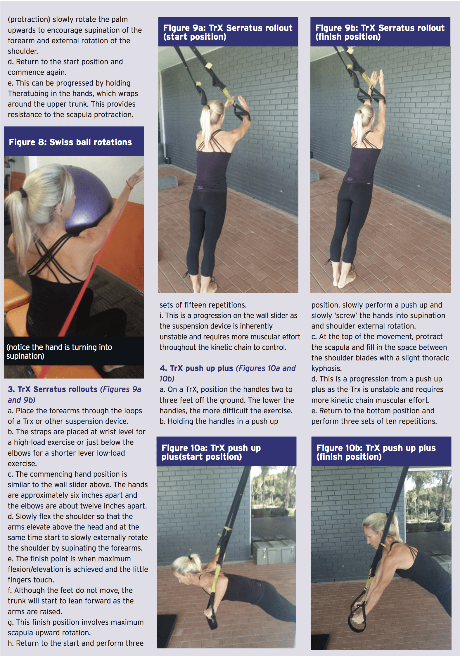
Exercises For SA
A significant amount of research has been conducted on finding the best rehabilitation exercises for the SA. The majority of these studies look at movements such as push ups, push up-plus exercises, cable and dumbbell ‘punch’ type movements. These exercises essentially emulate the function of the SA in its protraction role. Some of the findings of the more noted studies are;- Decker et al (1999) looked at the EMG activity and applied resistance associated with eight scapulohumeral exercises performed below shoulder height that target the SA muscle and how to design a continuum of SA muscle exercises for progressive rehabilitation or training(33). The best exercises according to these researchers are the push ups, dynamic hug, scaption and SA punch exercises.
- Barreto et al (2012) found high levels of activation of the SA in scaption exercises and adequate levels of activation of SA in MMP (modified military press)(34).
- Kim et al (2014) studied the interesting effect of vibration on SA activation and found that the push-up plus exercise performed using the Redcord system with mechanical vibration at 50 Hz increases SA muscle activity(35).
- Park and Yoo (2011) evaluated the effect of unstable surface on the upper and lower parts of the SA, while performing variations of the push-up exercise (push up and push up plus(36). The results indicated that the different parts of SA have distinct functions in the stabilisation process and therefore are recruited differently. The authors concluded that the main role of the lower SA is the fixation of the scapula onto the thoracic wall and therefore recommend performing the push-up plus on an unstable surface as a more effective strategy for the selective mobilisation of this component of the SA.
- Seo et al (2013) also found that the performance of a push up and knee push up on an unstable surface (Swiss ball) increased activation of the SA(37).
Below are some examples of clinically used SA activation exercises that anecdotally seem to recruit SA to high levels of function:

Summary
The serratus anterior (SA) is a muscle that plays an important role in the dynamic movement and control of the scapula during pushing movements and overhead elevation. Specifically it is a protractor, upward rotator, posterior tilt muscle of the scapula and additionally it fixes the scapula against the rib cage during movement. It is an important muscle for the overhead athlete as dysfunction in this muscle can lead to shoulder injuries such as impingement, rotator cuff breakdown and performance decrements during overhead tasks. This article has highlighted some clinically relevant exercises designed to retrain SA function in the athlete with SA dysfunction.References
1. J.Athl. Train. 2007; 42(2):311-319.
2. Phys Ther. 2000;80(3):276–291.
3. J OrthopSports Phys Ther 1999. 29: 574–586
4. J Orthop Sports Phys Ther 1996. 24: 57–65
5. J. Orthop. Sports Phys. Ther. 1994;20(6):307- 318.
6. Drake RL, Vogl W, Mitchell AWM. Gray’s anatomy for students. Philadelphia: Elsevier Inc; 2005. p. 633–47.
7. Clin Orthop Relat Res 1999. 368:17–27.
8. J BoneJoint Surg 1979;61:825–32
9. Simons et al (1999) Travell and Simons’ Myofascial Pain and Dysfunction. Volume 1 Upper Half of the Body (2nd edition). Williams and Wilkins. Baltimore.
10. J Bone Joint Surgery. 1944. 26(1); 1-30.
11. Am J Sports Med 2004; 32:1063–76.
12. J Orthop Sports Phys Ther 1993. 18: 427–432
13. Kibler WB: Normal shoulder mechanics and function. Instr Course Lect 46: 39–42, 1997
14. Am J Sports Med. 2003;31(4):542-9
15. Phys Ther 1994. 75: 194–202
16. Journal of orthopaedic & sports physical therapy. 2008. 38(1). 4-11
17. Contemp Orthop 1991. 22: 525–532
18. Physiol Ther. 2007;30(1):69-75
19. J Shoulder Elbow Surg 2000;9:31–5
20. J Shoudler Elbow Surg 1998;7:458–61
21. Curr Rev Musculoskelet Med 2008. 1:1–11
22. Chirurgie de la main 30 2011. 90–96
23. J. Bone & Joint Surg 1955. 37-A:567-574
24. Am J Sports Med 1991. 19: 569–576
25. J Bone Joint Surg 1988. 70A: 220–226
26. Am J Sports Med 1988. 16: 481–485
27. J Orthop Sports Phys Ther 1994. 20: 307–318
28. Am J Phys Med 1977;56(5):223–40
29. Am J Sports Med1991;19(6):577–82
30. Int J Sports Med 1997;18(8):618–24
31. J Electromyogr Kinesiol. 2005;15(6):576–586
32. Arch Phys Med Rehabil. 2002;83(1):60-9
33. Am J Sports Med 1999. Vol. 27, No. 6. 784-791
34. ConScientiae Saúde 2012. 11 (4) 660-667
35. J. Phys. Ther 2014. Sci. 26: 1275–1276
36. J Electromyogr Kinesiol. 2011 Oct;21(5):861-7
37. J Phys Ther Sci. 2013 Jul;25(7):833-7
